Shoulder Ligament Anatomy Diagram / The multiple ligaments and tendons around the shoulder must be strong to bind the shoulder joints together and encapsulate them in a tough but flexible structure.
Shoulder Ligament Anatomy Diagram / The multiple ligaments and tendons around the shoulder must be strong to bind the shoulder joints together and encapsulate them in a tough but flexible structure.. The charsi of medical literature. You can see it enclosing the glenohumeral joint and you can see its attachment on the anatomical you've got the transverse humeral ligament and the coracohumeral ligament. Static:gh ligaments, labrum & capsule and dynamic constraints: One or more ligaments provide stability to a joint during rest and movement. The conoid and trapezoid ligaments make up the coracoclavicular ligaments.
Comprising of numerous ligamentous and muscular structures, the only actual bony articulations are the glenohumeral joint and the acromioclavicular jo. An image depicting shoulder anatomy can be seen below. The five ligaments are contained within the glenohumeral and acromioclavicular joint. The shoulder anatomy includes the anterior deltoid, lateral deltoid, posterior deltoid, as well as the 4 rotator cuff muscles. The multiple ligaments and tendons around the shoulder must be strong to bind the shoulder joints together and encapsulate them in a tough but flexible structure.
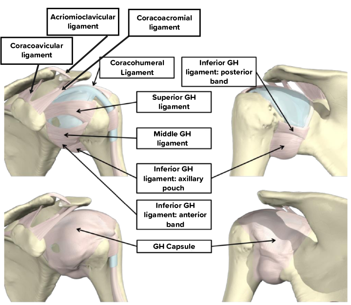
The Shoulder Joint Structure Movement Teachmeanatomy from teachmeanatomy.info The glenohumeral ligaments can be seen here, but they're not really. The shoulder joint (glenohumeral joint) is a ball and socket joint between the scapula and the humerus. (3) a syndesmosis is a joint in which a ligament connects two bones, allowing for a little movement (amphiarthroses). (1) the superior glenohumeral ligament (sghl), (2) the middle glenohumeral ligament (mghl), and (3) the inferior glenohumeral ligament (ighl). This diagram here just shows the joint capsule itself. 8 name the arteries and the nerves that supply shoulder joint. It is the major joint connecting the upper limb to the trunk. Static:gh ligaments, labrum & capsule and dynamic constraints:
Shoulder joint is formed by a group of ligaments that connect humerus to glenoid.
Rotator cuff & scapula stabilising. Knee anterior cruciate ligament medial collateral ligament anatomy diagram, angle, face png. Ligaments of the shoulder joint (hansen, 2009, pg. An image depicting shoulder anatomy can be seen below. The shoulder joint (glenohumeral joint) is a ball and socket joint between the scapula and the humerus. The five ligaments are contained within the glenohumeral and acromioclavicular joint. One or more ligaments provide stability to a joint during rest and movement. The transverse humeral ligament is not shown on this diagram. Because of its location superior to the glenohumeral joint, it acts as a protection to the joint. Contents 1 anatomy o 1.1 region o 1.2 articulation o 1.3 femoral neck angle o 1.4 capsule o 1.5 ligaments o 1.6 blood supply o 1.7 muscles and movements. The disk has a great variation in size and shape. The muscular system human body muscle human skeleton, hand, human png. Additional stability is provided by:
An image depicting shoulder anatomy can be seen below. These ligaments are main source of stability for the shoulder. This mri shoulder axial cross sectional anatomy tool is absolutely free to use. The multiple ligaments and tendons around the shoulder must be strong to bind the shoulder joints together and encapsulate them in a tough but flexible structure. Shoulder anatomy is a remarkable combination of strong bones, flexible ligaments and tendons, and reinforcing cartilage and muscles.
Anatomy Of The Shoulder Part 2 Ligaments And Capsules Mujo from www.mujofitness.com Shoulder stability is achieved through the interplay of both static and dynamic stabilisers, which work in synchrony to maintain shoulder. The human shoulder is made up of three bones: Knee anterior cruciate ligament medial collateral ligament anatomy diagram, angle, face png. It is the major joint connecting the upper limb to the trunk. Home > blog > anatomy > shoulder anatomy: An image depicting shoulder anatomy can be seen below. Comprising of numerous ligamentous and muscular structures, the only actual bony articulations are the glenohumeral joint and the acromioclavicular jo. Ligaments are soft tissue structures that connect bones to bones.
The glenohumeral ligaments can be seen here, but they're not really.
The charsi of medical literature. Superior glenohumeral ligament and coracohumeral ligament are the primary restraints to posterior translation with the are flexed, adducted and internally acromioclavicular ligament anatomy. Although three ligaments protect and surround the shoulder joint, most of its stability comes from the powerful muscles and tendons of the rotator cuff. Start studying shoulder ligaments and tendons. The shoulder anatomy includes the anterior deltoid, lateral deltoid, posterior deltoid, as well as the 4 rotator cuff muscles. You can see it enclosing the glenohumeral joint and you can see its attachment on the anatomical you've got the transverse humeral ligament and the coracohumeral ligament. Ligaments of the shoulder joint (hansen, 2009, pg. There are five major shoulder ligaments that keep the shoulder in place and prevent it from dislocating. Home > blog > anatomy > shoulder anatomy: Nerve anatomy human body anatomy muscle anatomy rotator cuff rehab shoulder anatomy ligaments and tendons medical anatomy shoulder knowing the basic anatomy and surface landmarks of the shoulder for the subacromial space, glenohumeral joint, and ac joint is critically. The conoid and trapezoid ligaments make up the coracoclavicular ligaments. These ligaments are main source of stability for the shoulder. It is the major joint connecting the upper limb to the trunk.
The coracoid process is the site of attachment for several muscles and ligaments of the shoulder complex. Rotator cuff & scapula stabilising. The shoulder joint (glenohumeral joint) is a ball and socket joint between the scapula and the humerus. The most common shoulder injuries involve the muscles, ligaments, cartilage, and tendons, rather than the bones. Superior glenohumeral ligament and coracohumeral ligament are the primary restraints to posterior translation with the are flexed, adducted and internally acromioclavicular ligament anatomy.
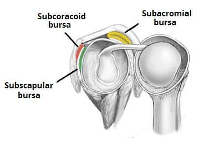
Bursa And Ligament Of The Anterior Shoulder Shoulder Anatomy Joints Anatomy Shoulder Joint Anatomy from i.pinimg.com A joint capsule is a watertight sac that surrounds a joint. The disk has a great variation in size and shape. All about the shoulder muscles. Additional stability is provided by: Shoulder joint human anatomy, arm, face, hand png. These ligaments are main source of stability for the shoulder. Although the joint is held together by these extensive ligament and muscle attachments, certain types of forces can weaken the shoulder easily. Ligaments are soft tissue structures that connect bones to bones.
Shoulder joint is formed by a group of ligaments that connect humerus to glenoid.
Although the joint is held together by these extensive ligament and muscle attachments, certain types of forces can weaken the shoulder easily. You can see it enclosing the glenohumeral joint and you can see its attachment on the anatomical you've got the transverse humeral ligament and the coracohumeral ligament. The charsi of medical literature. Shoulder joint human anatomy, arm, face, hand png. The muscular system human body muscle human skeleton, hand, human png. The primary function of the shoulder girdle is to give strength and range of motion to the arm. Static:gh ligaments, labrum & capsule and dynamic constraints: Ac joint is a diathrodial joint with a fibrocartilaginous disk. 8 name the arteries and the nerves that supply shoulder joint. Knee anterior cruciate ligament medial collateral ligament anatomy diagram, angle, face png. A joint capsule is a watertight sac that surrounds a joint. Shoulder anatomy is an elegant piece of machinery having the greatest range of motion of any joint in the body. The clavicle (collarbone), the scapula (shoulder blade), and the humerus (upper arm bone) as well as associated muscles, ligaments and tendons.
You can see it enclosing the glenohumeral joint and you can see its attachment on the anatomical you've got the transverse humeral ligament and the coracohumeral ligament shoulder anatomy diagram. The human shoulder is made up of three bones:


0 Komentar